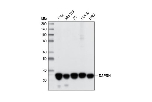

Detection reagent used: m-IgG BP-CFL 555: sc-516177. Western blotting (WB) or immunoblotting is a widely used laboratory method to detect multiple epitopes and semiquantify tissue proteins levels. Blocked with UltraCruz ® Blocking Reagent: sc-516214.
#Gapdh western blot Activator#
Signal transducer and activator of transcription 3 inhibition alleviates resistance to BRAF inhibition in anaplastic thyroid cancer. Fluorescent western blot analysis of GAPDH expression in MCF7 (A), HeLa (B), C32 (C), Jurkat (D), Hep G2 (E) and BJAB (F) whole cell lysates. Am J Physiol Gastrointest Liver Physiol 320:G816-G828 (2021). Long noncoding RNA LINC00982 upregulates CTSF expression to inhibit gastric cancer progression via the transcription factor HEY1. Preventing oxaliplatin-induced neuropathic pain: Using berberine to inhibit the activation of NF-?B and release of pro-inflammatory cytokines in dorsal root ganglions in rats. Upregulated microRNA-126 induces apoptosis of dental pulp stem cell via mediating PTEN-regulated Akt activation. GATA binding protein 5-mediated transcriptional activation of transmembrane protein 100 suppresses cell proliferation, migration and epithelial-to-mesenchymal transition in prostate cancer DU145 cells. Though differentially expressed from tissue to tissue, GAPDH is frequently used as a loading control for assays involving mRNA and protein detection. Publishing research using ab9485? Please let us know so that we can cite the reference in this datasheet.Īb9485 has been referenced in 2293 publications. Antibody binding was detected using Goat Anti-Rabbit IgG H&L (Alexa Fluor® 790) (ab175781) secondary antibody at a 1:10,000 dilution for 1hr at room temperature and then imaged using the Licor Odyssey CLx. The membrane was then blocked for an hour using Licor blocking buffer before being incubated with ab9485 overnight at 4☌. The gel was run at 200V for 50 minutes before being transferred onto a Nitrocellulose membrane at 30V for 70 minutes.

This blot was produced using a 4-12% Bis-tris gel under the MOPS buffer system. GAPDH is constitutively expressed at high levels in almost all. Lane 3 : A549 (Human lung adenocarcinoma epithelial cell line) Whole Cell LysateĪll lanes : Goat Anti-Rabbit IgG H&L (Alexa Fluor® 790) ( ab175781) secondary antibody at 1/10000 dilution GAPDH loading control antibody is ideal for Western Blotting, ELISA, IHC and IF Microscopy. Lane 2 : A431 (Human epithelial carcinoma cell line) Whole Cell Lysate Lane 1 : HeLa (Human epithelial carcinoma cell line) Whole Cell Lysate All lanes : Anti-GAPDH antibody - Loading Control (ab9485) at 1/2500 dilution


 0 kommentar(er)
